Neumann Lab
  • Home
  • Our Research
    • Axonal Regeneration
    • Charcot-Marie-Tooth Disease and Mfn2
    • Axonal Degeneration
  • Our Team
    • Dr Brent Neumann
    • Jean-Sebastien Teoh
    • Tarika Vijayaraghavan
    • Samiksha Dhananjay
  • Publications
  • News/Blog
  • Image gallery
  • Contact
  • Links

Our first major foray into understanding CMT

16/4/2020

0 Comments

 
We have published our first major study on Charcot-Marie-Tooth disease (CMT) to the bioRxiv preprint server (see our publications page for links).

CMT is divided into demyelinating (CMT1) and axonal (CMT2) neuropathies, and although we have gained molecular information into the details of CMT1 pathology, much less is known about CMT2. Mutations in more than 20 genes associated with diverse cellular functions are known to be causative for different subtypes of CMT2. Thus, a major unresolved question in the field is how mutations affecting seemingly functionally distinct proteins all lead to the same symptoms in CMT patients. In an attempt to decipher the biological roles of the genes known to cause CMT2, we have targeted nine orthologous genes in the nematode C. elegans and performed comprehensive analyses into the consequences on animal behaviour and cellular/molecular functioning.

Our study reveals common deficiencies in muscle architecture and function, and consequent locomotion difficulties amongst these genetic mutants. Furthermore, our study reveals deterioration of cholinergic neuronal structure as a result of CMT2-related genetic dysfunction, and prevailing deficits at the neuromuscular junction in animals presenting the most severe phenotypes.

We provide the first non-subjective, quantitative methods for assessing muscle morphology in C. elegans. Our methods can help move the field away from subjective, qualitative methods and towards reliable, unbiased quantitative assessments not only in the muscles, but other cell types as well. We also describe detailed methodology for the semi-automated analysis of C. elegans locomotion in populations of animals grown in liquid culture conditions. We use these procedures to demonstrate evident differences between wild-type and animals lacking CMT2-associated genes that will provide excellent readouts for future high-throughput drug screens to identify effective therapeutics that can reverse the locomotion defects. 

Finally, our results greatly expand our knowledge into the cellular roles for nine different CMT2-related genes. The biological roles for many of the causative gene have been poorly described. By simultaneously assessing the effects of mutating nine different causative genes in a whole animal model, we have been able to compare deficiencies across CMT2 subtypes. This has revealed that each is essential for robust muscle structure, and importantly that most are involved in cholinergic neurotransmission and maintaining functional neuromuscular junctions. Thus, our results identify common cellular defects when these genes are disrupted, and suggest that the neuromuscular junction could be a potential therapeutic target for CMT2.
0 Comments

Paper Published in FASEB J!!

1/12/2019

0 Comments

 
Our manuscript entitled "Axonal repair by fusion: pitfalls, consequences and solutions" has been published as a perspective article in The FASEB Journal!

Given the clinical challenges associated with repairing damaged nerves, using model organisms to define the molecular mechanisms controlling spontaneous modes of functional regeneration can be advantageous. In the last five years, using the nematode Caenorhabditis elegans as a model system, we have gained major insights into the molecular control of a highly efficient repair paradigm known as axonal fusion.  In this key biological process, individually transected axons regrow, reconnect, and fuse, with their own separated distal axon fragments, restoring the original axonal tract and the full neuronal function. Excitingly, similar paradigms that have been optimised in rodent and canine models have now been successfully used in the clinic.
 
However, based on the current literature and from conversations with colleagues, it has become apparent that there are conceptual differences in the way axonal fusion is analysed and quantified, potentially leading to the publication of misleading or incorrect findings. Thus, it is essential that a unifying view is developed to clarify how to precisely examine and score this biological event in order to prevent the publication and dissemination of erroneous interpretations. In our manuscript, we address this by providing clear examples of how fusion should be assessed and the errors that can result from incorrect analysis. We hope that our article will be a helpful guide as more groups focus on regenerative axonal fusion.

See here for a link to our manuscript.
0 Comments

Congratulations Tarika!!

22/11/2019

0 Comments

 
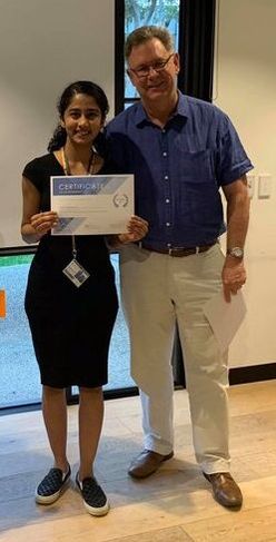
Tarika has been awarded the runner up prize for best oral presentation at the Anatomy and Developmental Biology Postgraduate Student Symposium!!

Her presentation was entitled "The Dynamin GTPase is required for regeneration axonal fusion".

Well done Tarika!!
Picture
Tarika with our Department head John Bertram.
0 Comments

Australian C. elegans Symposium 2019

29/10/2019

0 Comments

 
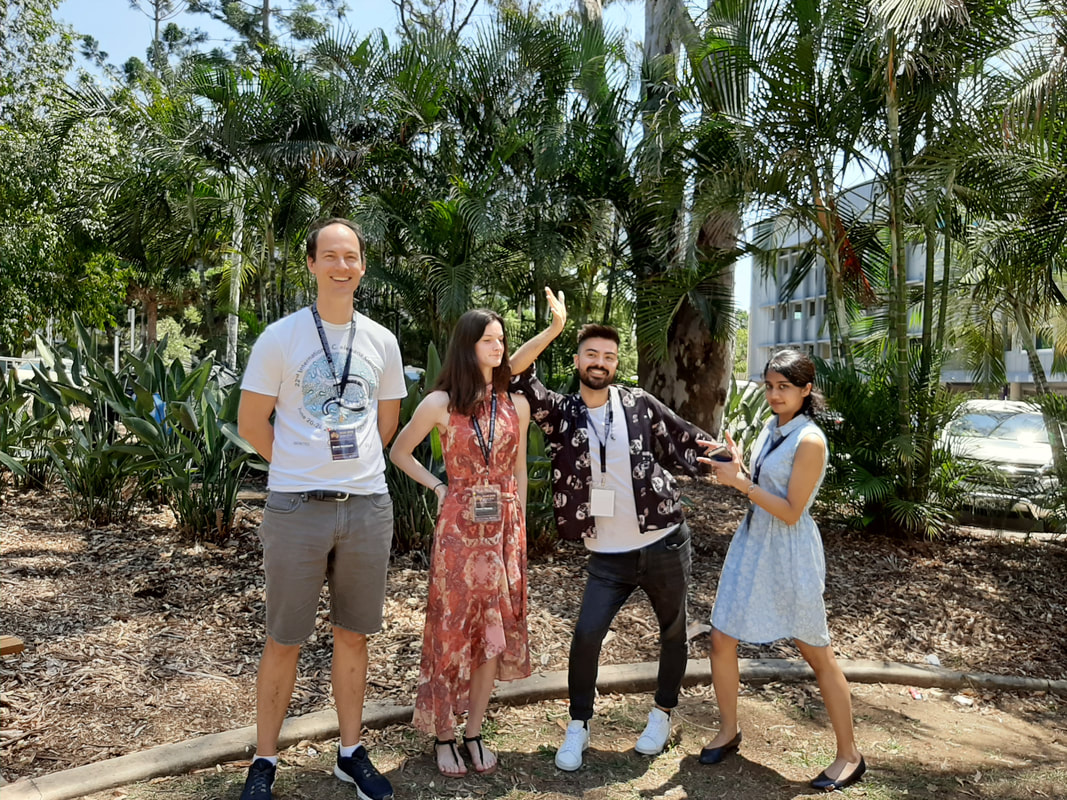
Seb, Tarika, Arwen and Brent all attended the second Australian C. elegans Symposium (ACeS) held at the Queensland Brain Institute in sunny Brisbane.

Seb presented a talk entitled: The α-tubulin acetyltransferase MEC-17/αTAT1 is essential for robust axonal regeneration.
Tarika presented a talk entitled: Role of Dynamin GTPases in axonal fusion.
Arwen presented a poster entitled: Characterisation of novel genes involved in mitochondrial fusion.
Brent was part of the organising committee.

Check out some photos of the meeting here:
​

More information of the conference can be found here:
https://www.australianceleganssymposium.org/
Picture
0 Comments

Neumann Lab vs Art

3/10/2019

0 Comments

 
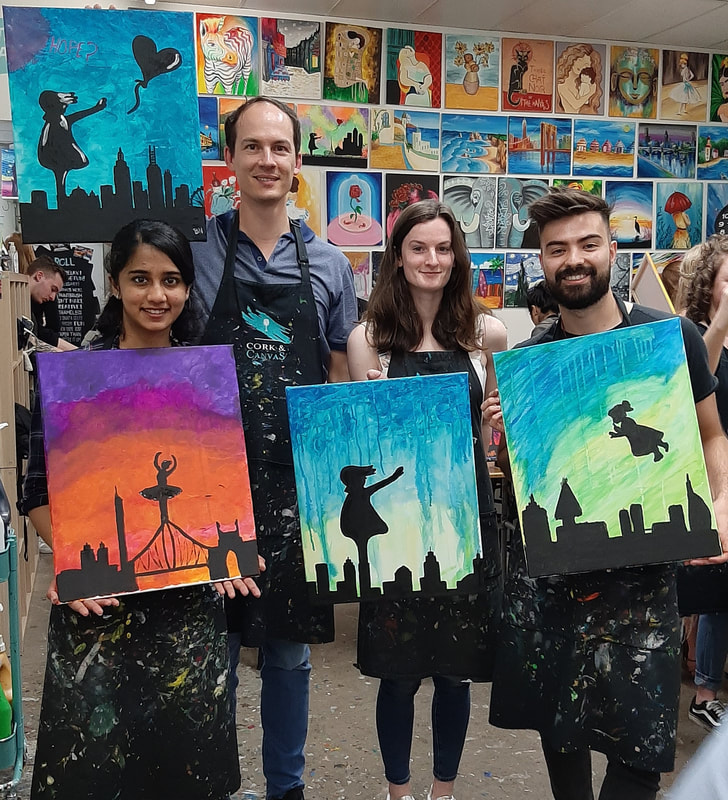
We had a night out at Corks and Canvas at South Melbourne creating some Banksy-inspired masterpieces.

Great work everyone!

​See more pics here.
Picture
0 Comments

Paper Published in JOVE

6/7/2019

0 Comments

 
We have had a manuscript accepted for publication by the Journal of Visualized Experiments. This paper is a co-first author publication for Seb, Ming and Joe - congratulations to all three!!

Our paper is titled "Quantitative Approaches for Studying Cellular Structures and Organelle Morphology in Caenorhabditis elegans".

In this manuscript, we outline quantitative methods for the analysis of synaptic size and integrity, muscle morphology, and mitochondrial shape in C. elegans using freely available image processing tools. 

Many studies in C. elegans use qualitative methods for comparing differences in the morphology of organelles and tissues. However, these can be problematic as they may not capture subtle phenotypic differences, might under- or over-represent variations across individuals in a population, and are assessed subjectively. We have developed quantitative methods using a number of different freely available programs (Fiji, ilastik, CellProfiler, SQUASSH) to assess the morphology of synapses, muscles and mitochondria. We believe that these methods provide more robust and less biased means for assessing morphological changes as a result of genetic mutations or changes in environmental conditions.

Our quantitative approaches are not limited to the applications we describe, as they could readily be used to assess the morphology of other tissues and organelles in the nematode, as well as in other model organisms or in cultured cells. We hope that our techniques will help to shift the field away from categorical assessments and towards non-subjective quantifications.
0 Comments

Paper featured on the cover of CMLS

26/4/2019

0 Comments

 
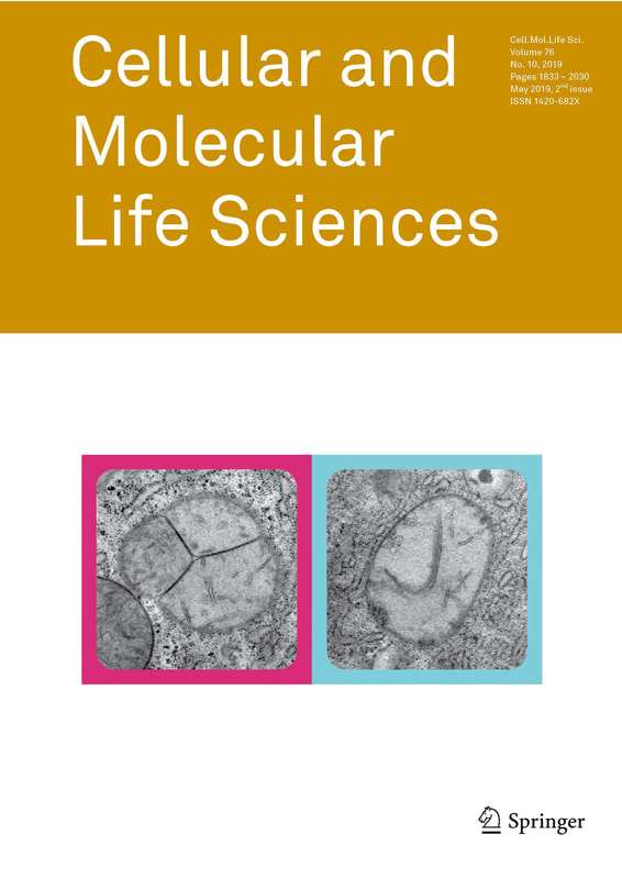
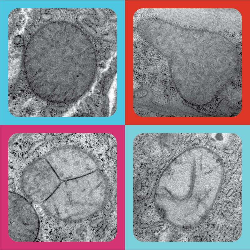
Our paper in Cellular and Molecular Life Sciences on the role of mitochondrial fission and fusion proteins in C. elegans behaviour and lifespan has been featured on the cover of the journal!

The image shows the ultrastructure of mitochondria within the body wall muscles of C. elegans lacking either of the major mitochondrial fusion proteins (FZO-1 and EAT-3). The image was submitted as a four panel collage (see image at bottom) for consideration, but a two panel version was requested by the journal (see below - still looks great!).
Picture
Our image on the cover of the journal!
Picture
The original pop-art inspired four-panel image that we submitted
0 Comments

Lab Trophy

19/4/2019

0 Comments

 
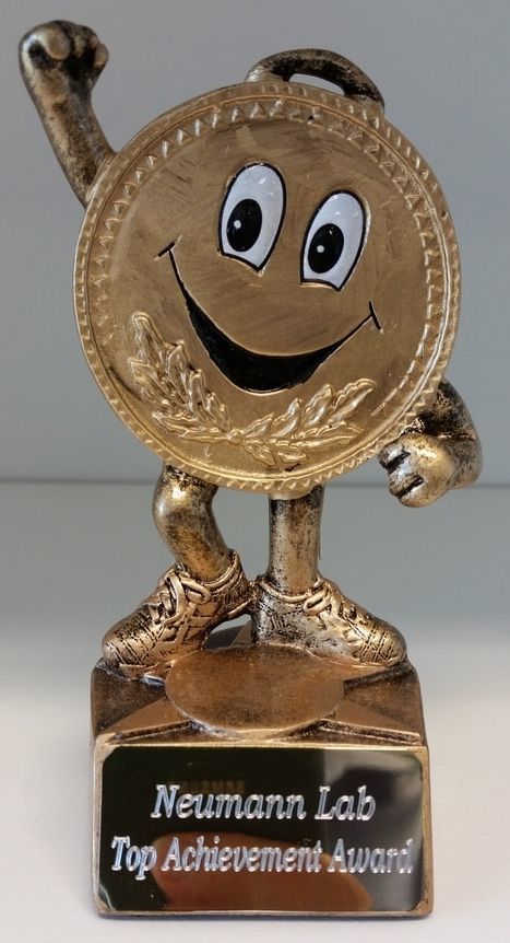
The highly prestigious Neumann Lab trophy is awarded to a member of the lab who has achieved great success or for any other reason we deem appropriate!

The inaugural recipient was Valerie for getting strain number 100 - a wonderfully random achievement!

Current holder of the Top Achievement Award:

​Michelle: Strain M for Michelle 


Awarded for adding the 1000th C. elegans strain to our lab collection.

Honour board
11. Michelle: Strain M for Michelle - adding the 1000th strain to the lab collection.
10. Simran: What the HEK! - culturing mammalian cells (HEK293T's) for the first time in the                          Neumann lab.
  9. Tarika: CRISPR Queen - successfully getting a vital CRISPR mutant.
  8. Ming: Drug Tester - completing a pilot drug screen to identify effective therapeutics for                            Charcot-Marie-Tooth disease.
  7. Ming: Magnificent Muscles - taking a truly beautiful image of the C. elegans body wall                            muscles (now on our CMT research page - see here).
  6.  Michelle: Confirmed! - successfully completing her 12 month requirements as a PhD                              candidate.
  5.  Tarika: Money Bags - being awarded three seperate scholarships on a single day.
  4.  Michelle: WGS Success - successful sequencing and mapping various mutants isolated                        from genetic screening.
  3.  Grace: First lab thesis - the first thesis completed in the Neumann Lab.
  2.  Joe: CRISPR King - successfully getting a CRISPR mutant for the first time in the lab.
  1.  Valerie: Strain 100 - generating strain #100 in the Neumann Lab.
0 Comments

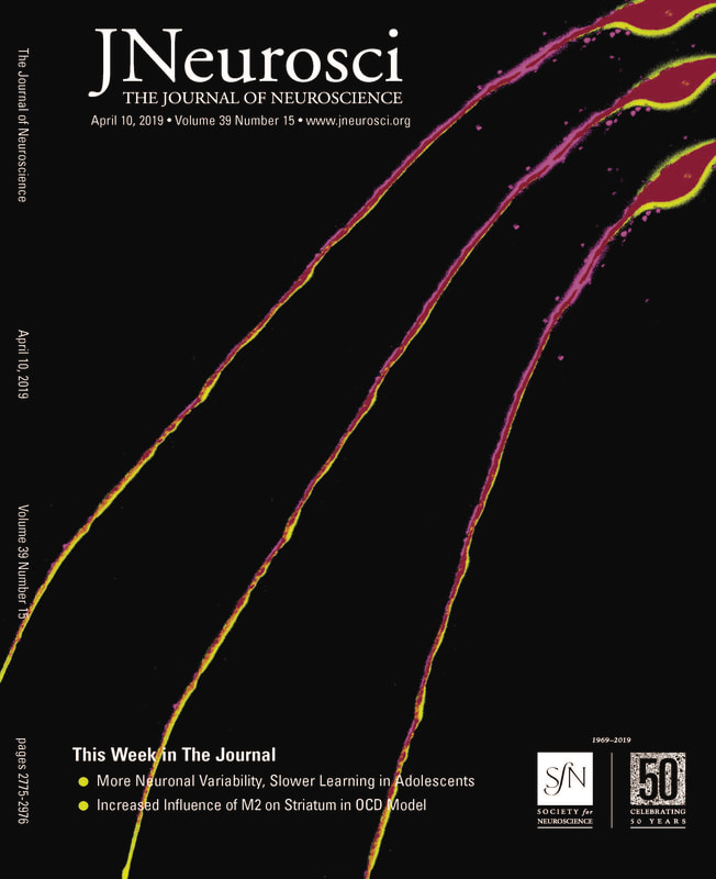
paper featured on the cover of J Neuroscience!

16/4/2019

0 Comments

 
Our paper in the Journal of Neuroscience has been featured on the cover of the journal!

The cover can be seen below and here: ​http://www.jneurosci.org/
Picture
0 Comments

Tarika publishes in Cell Reports!!

27/3/2019

0 Comments

 
Research from Tarika's Masters project in Arnim Pause's lab has been published in Cell Reports!!

Congratulations Tarika!

The paper can be found here.
0 Comments
<<Previous

    Categories

    All
    Awards
    Blog
    Events
    News
    Publication Info
    Trophy

    Picture

    Archives

    April 2020
    November 2019
    October 2019
    July 2019
    April 2019
    March 2019
    January 2019
    December 2018
    November 2018
    October 2018
    September 2018
    August 2018
    July 2018
    June 2018
    May 2018
    April 2018
    February 2018
    December 2017
    November 2017
    October 2017
    September 2017
    August 2017
    July 2017
    June 2017
    May 2017
    April 2017
    March 2017
    February 2017
    January 2017
    December 2016
    November 2016
    October 2016
    August 2016
    June 2016
    May 2016
    April 2016
    March 2016
    February 2016
    January 2016
    December 2015
    November 2015
    October 2015
    September 2015

    RSS Feed

Our Research

Axonal Regeneration
Charcot-Marie-Tooth Disease & Mfn2
Axonal Degeneration

Our Team

Lab members
Contact
Publications

Lab News

​Latest news.........
Paper accepted in PNAS Nexus!!
Follow @neumannlab
Biomedicine Discovery Institute
© COPYRIGHT 2015. ​ALL RIGHTS RESERVED.